Stages of Endometrial Cancer

staging-endometrial-cancer

If endometrial cancer is diagnosed, you will be referred to a gynecologic oncologist — the specialist who will lead your cancer treatment, including surgery to remove the uterus. The gynecologic oncologist will also do an evaluation during surgery to determine if the cancer has spread to other areas of the body.

Tests Used to See if Cancer has Spread

When the oncologist removes the uterus, some nearby lymph nodes and tissue samples from the pelvis and abdomen are usually removed and checked to see how deeply the cancer has spread.

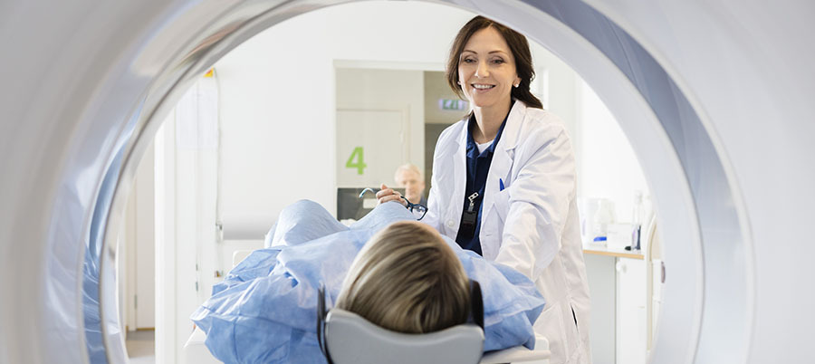
Sometimes, the gynecologic oncologist may need to conduct other tests in order to be sure they know exactly where the cancer is located in the body. One or more of the following tests may be requested:

  • Chest X-ray: An x-ray of the organs and bones inside the chest can show a tumor in the lung.

  • PET scan (positron emission tomography scan): Combined with a CT Scan, this type of scan will find other malignant tumor cells in the body. This is done by giving the patient a small amount of radioactive glucose (sugar) so that cancerous cells in the body will “light up” and be visible on the scan, even if they aren’t visible to the human eye.

  • MRI (magnetic resonance imaging): A large machine with a strong magnet linked to a computer is used to make detailed pictures of your uterus and lymph nodes.

  • Lymph node biopsy: A surgical procedure in which a few lymph nodes are removed from the pelvic area and a sample of tissue is checked under a microscope for signs of cancer. It can be done before surgery to remove the cancer, but is often done at the same time.

New call-to-action

Stages of Endometrial Cancer

These are the stages of endometrial cancer classified by The International Federation of Gynecology and Obstetrics. You may also be given a substage that indicates how far the cancer has spread within that stage.

  • Stage I: The tumor has grown through the inner lining of the uterus to the endometrium. It may have invaded the myometrium.

  • Stage II: The tumor has invaded the cervix.

  • Stage III: The tumor has grown through the uterus to reach nearby tissues, such as the vagina or a lymph node.

  • Stage IV: The tumor has invaded the bladder or intestine, or cancer cells have spread to parts of the body far away from the uterus, such as the liver, lungs or bones.

When cancer spreads from its original place to another part of the body, the new tumor has the same kind of abnormal cells and the same name as the primary (original) tumor. For example, if endometrial cancer spreads to the ovaries, the cancer cells on the ovaries are actually endometrial cancer cells and would be treated as such. Doctors sometimes call the new tumor “distant” disease.

What is the Grade of Cancer?

If cancer is found, the pathologist studies tissue samples from the uterus under a microscope to learn the grade of the tumor. The grade tells how much the tumor tissue differs from normal uterine tissue. It may suggest how fast the tumor is likely to grow. Tumors with higher grades (called “poorly differentiated”) tend to grow faster than those with lower grades (called “differentiated”). Tumors with higher grades are also more likely to spread. Doctors use tumor grade along with other factors to suggest treatment options. In general, the slower the spread, the better the prognosis.

The letter "G" is used to define a grade for endometrial cancer.

  • GX: The grade cannot be evaluated.

  • G1: The cells are well differentiated, meaning they aren’t too different from normal cells. This might be referred to as low grade.

  • G2: The cells are moderately differentiated.

  • G3: The cancer cells look very different from healthy cells. This might also be referred to as high grade.