
Doctors use a variety of tests, procedures, and scans to examine the throat and neck in order to detect and diagnose hypopharyngeal cancer. If you exhibit signs and symptoms of head and neck cancer, the doctor will take a complete medical history, noting all symptoms and risk factors. He or she may also refer you to an Ear, Nose, Throat specialist (ENT) who will perform a more complete exam and request further testing, such as biopsy and imaging if necessary.
Your PCP or ENT will evaluate your signs and symptoms with:
Physical exam and health history: A simple exam of the body to check general signs of health, including checking for signs of disease, such as lumps or anything else that seems unusual. A history of the patient’s health habits and past illnesses and treatments will also be taken. Blood tests and urine tests may be recommended.
If referred to an ENT for further examination, some of the following tests for procedures may be run to make a determination:
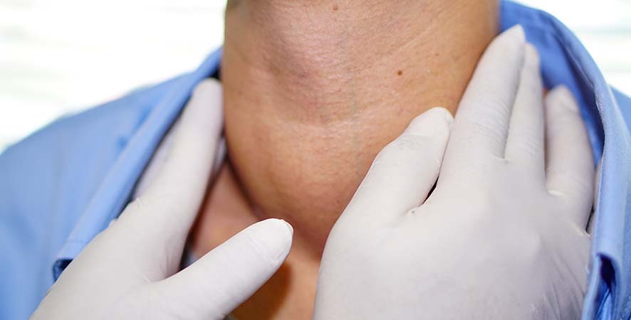
Physical exam of the throat: A doctor will feel for swollen lymph nodes in the neck and look down the throat with a small, long-handled mirror to check for areas of concern.
Neurological exam: Multiple questions and tests to check the brain, spinal cord, and nerve function. The exam analyzes a person’s mental status, coordination, and ability to walk normally, and how well the muscles, senses, and reflexes work.
Endoscopy: A procedure used to look at areas in the throat that cannot be seen with a mirror during the physical exam of the throat. An endoscope (a thin, lighted tube) is inserted through the nose or mouth to check the throat for anything that seems unusual. Tissue samples may be taken for biopsy.
Laryngoscopy: A procedure to look at the larynx (voice box) for abnormal areas. A mirror or a laryngoscope (a thin, tube-like instrument with a light and a lens for viewing) is inserted through the mouth to see the larynx.
Panendoscopy: a procedure that involves a combination of laryngoscopy, esophagoscopy, and in some cases, bronchoscopy. A panendoscopy is a test used to look at the pharynx, larynx, upper trachea, and esophagus.
Videostroboscopy: A technique that uses fiber optics to enhance the view of the larynx in order to detect motion abnormalities and other changes in vibration within the vocal folds. Videostroboscopy helps determine the location and size of a tumor, as well as how the tumor has affected the function of the larynx and hypopharynx. This method is useful because the results often show potential changes before they are visible to the eye alone.
If cancer is suspected, the doctor may recommend a biopsy. While other tests can suggest that cancer is present, only a biopsy can make a definite diagnosis. A biopsy is the removal of cells or tissues so they can be viewed under a microscope to check for signs of cancer. The specialist can take out small tissue samples from any tumors or other changed areas using special tools put through an endoscope, laryngoscope, or bronchoscope.
If the biopsy confirms cancer, it’s common to run additional tests including blood work and imaging, to see if the cancer has spread and, if so, how far. Your doctor may request imaging tests including:
CT scan (CAT scan): A procedure that makes a series of detailed pictures of areas inside the body, such as the head, neck, chest, and lymph nodes, taken from different angles. The pictures are made by a computer linked to an x-ray machine. A dye may be injected into a vein or swallowed to help the organs or tissues show up more clearly. This procedure is also called computed tomography, computerized tomography, or computerized axial tomography.
PET scan (positron emission tomography scan): A procedure to find malignant (cancerous) tumor cells in the body. A small amount of radioactive glucose (sugar) is injected into a vein. The PET scanner rotates around the body and makes a picture of where glucose is being used in the body. Malignant tumor cells show up brighter in the picture because they are more active and take up more glucose than normal cells do. A PET scan and CT scan may be done at the same time. This is called a PET-CT.
MRI (magnetic resonance imaging): A procedure that uses a magnet, radio waves, and a computer to make a series of detailed pictures of areas inside the body. This procedure is also called nuclear magnetic resonance imaging (NMRI).
Bone scan: A procedure to check if there are rapidly dividing cells, such as cancer cells, in the bone. A very small amount of radioactive material is injected into a vein and travels through the bloodstream. The radioactive material collects in the bones with cancer and is detected by a scanner.
Barium esophagogram: An x-ray of the esophagus. The patient drinks a liquid that contains a silver-white metallic compound called barium. The liquid coats the esophagus and x-rays are taken.
Quickly and efficiently build the materials you need to support your inbound marketing strategy. Drag and drop building blocks including testimonials, forms, calls-to-action, and more.