
If your doctor suspects oral cancer, he or she may do further examinations and perform some tests to see if there are signs that cancer is developing.
Initial evaluation of the lips and mouth when oral cancer is suspected include:
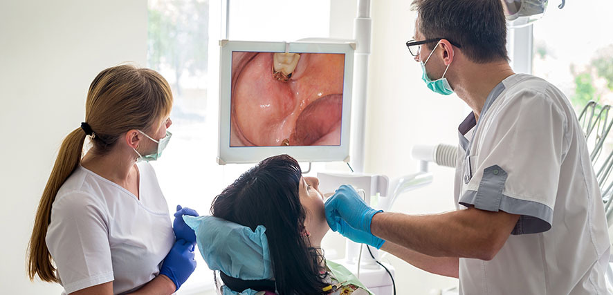
Physical exam of the lips and oral cavity: An exam to check the lips and oral cavity for abnormal areas. This will include checking the insides of the cheeks and lips, the gums, the roof and floor of the mouth, and the top, bottom, and sides of the tongue. The neck will be felt for swollen lymph nodes. A history of the patient’s health habits and past illnesses and medical and dental treatments will also be taken.
Exfoliative cytology: A procedure to collect cells from the lip or oral cavity. A piece of cotton, a brush, or a small wooden stick is used to gently scrape cells from the lips, tongue, mouth, or throat. The cells are viewed under a microscope to find out if they are abnormal. This can be done in the dentist’s office very simply. If something is found in the results, further testing is recommended such as a biopsy.
Endoscopy: Using an endoscope — a thin, tube-like instrument with a light and a lens— the doctor can look inside the mouth and throat. This is especially helpful for seeing down the throat for anything that looks unusual and should be biopsied.
If cancer is suspected based on initial tests, further testing is likely. This is to determine if the cells are in fact cancerous, and to see if the cancer has spread beyond the oral cavity. These tests include the following, although not all of these tests will necessarily be done right away.
Biopsy: The removal of cells or tissues so they can be viewed under a microscope by a pathologist.
HPV test: This is done on cells gathered in the biopsy to help the doctor know if the human papilloma virus is present. If it is, the recommended treatments may be adjusted.
MRI (magnetic resonance imaging): A procedure that uses a magnet, radio waves, and a computer to make a series of detailed pictures of areas inside the body. This procedure is also called nuclear magnetic resonance imaging (NMRI).
CT scan (CAT scan): A procedure that makes a series of detailed pictures of areas inside the body, taken from different angles. The pictures are made by a computer linked to an x-ray machine. A dye may be injected into a vein or swallowed to help the organs or tissues show up more clearly.
Barium swallow: A series of x-rays of the esophagus and stomach. The patient drinks a liquid that contains barium (a silver-white metallic compound). The liquid coats the esophagus and x-rays are taken. This procedure is also called an upper GI series.
PET scan (positron emission tomography scan): A procedure to find malignant tumor cells in the body. A small amount of radioactive glucose (sugar) is injected into a vein. The PET scanner rotates around the body and makes a picture of where glucose is being used in the body.
Bone scan: A procedure to check if there are rapidly dividing cells, such as cancer cells, in the bone. A very small amount of radioactive material is injected into a vein and travels through the bloodstream. The radioactive material collects in the bones with cancer and is detected by a scanner.